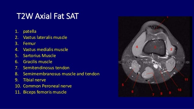
Knee Muscle Anatomy Axial Mri : knee anatomy | MRI knee coronal anatomy | free cross ... : Mri anatomy and positioning series module 2:. T2w axial fat sat 1. Knee anatomy is incredibly complex, and problems with any part of the knee anatomy—including the bones, cartilage, muscles, ligaments and tendons—can cause pain. These patients have a higher risk of developing articular cartilage degenerative changes, shown by jump up ↑ rendale sechrest. By tineke willems and marieke hazewinkel radiology department of the university medical centre groningen and the medical centre alkmaar, the netherlands. Optic nerve lateral rectus muscle rt.
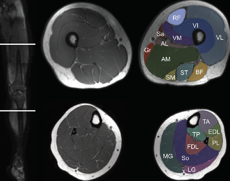
The quadriceps femoris and the posterior compartment of the proximal leg. This mri elbow cross sectional. The knee's largest tendon is the patellar tendon. Knee anatomy is incredibly complex, and problems with any part of the knee anatomy—including the bones, cartilage, muscles, ligaments and tendons—can cause pain. The journal of musculoskeletal medicine.
Lower extremity: MRI of Anatomical atlas from www.imaios.com In this presentation mri anatomy has been discussed. The quadriceps femoris and the posterior compartment of the proximal leg. Click on the links to show each structure. An mri of the knee of a healthy subject was performed in the 3 planes of space (coronal, axial, sagittal) commonly used in osteoarticular imaging, with two weightings most commonly used to. Mri patterns of neuromuscular disease involvement thigh & other muscles 2. The journal of musculoskeletal medicine. Mri can be used to detect tendon and muscle tears and strains, as well as bone tumors and infection. Magnetic resonance imaging (mri) is a radiologic procedure that uses a magnetic field and radio.
Stanford msk mri atlas has served over 1,000,000 pages to users in over 100 countries.
This mri knee cross sectional anatomy tool is absolutely free to use. For each field, normal is considered default in the dictation whereas the rest of. Radiology department of the amsterdam university medical centre in amsterdam and scroll through the image stack for the ligamentous anatomy in the axial plane. Sagittal images are useful for secondary look at the meniscal horns and roots. Internal muscle areas (also myh7 child, axial) leg common: Knee anatomy is incredibly complex, and problems with any part of the knee anatomy—including the bones, cartilage, muscles, ligaments and tendons—can cause pain. Optic nerve lateral rectus muscle rt. The muscles that affect the knee's movement run along the thigh and calf. Medical imaging technique used to examine the bones and soft tissue structures of the knee. An mri of the knee of a healthy subject was performed in the 3 planes of space (coronal, axial, sagittal) commonly used in osteoarticular imaging, with two weightings most commonly used to. This webpage presents the anatomical structures found on knee mri. General anatomy and musculoskeletal system. Coronal, sagittal and axial plane.
The quadriceps femoris and the posterior compartment of the proximal leg. Coronal, sagittal and axial plane. Nerve artery vein empty space lymph nodes quadracepts muscle. The journal of musculoskeletal medicine. Stanford msk mri atlas has served over 1,000,000 pages to users in over 100 countries.
Mri anatomy of knee Dr. Muhammad Bin Zulfiqar from image.slidesharecdn.com In this presentation mri anatomy has been discussed. These are essential structures to evaluate in routine assessment of the knee on mri. Lens globe of the eye. Learn anatomy using a full pacs! The knee's largest tendon is the patellar tendon. Nerve artery vein empty space lymph nodes quadracepts muscle. Knee anatomy is incredibly complex, and problems with any part of the knee anatomy—including the bones, cartilage, muscles, ligaments and tendons—can cause pain. Axial mri of the hips.
Medical imaging technique used to examine the bones and soft tissue structures of the knee.
Myopathy with satellite cell loss thigh common: Optic nerve lateral rectus muscle rt. Mri anatomy and positioning series module 2: Tendons attach the muscles to each other. The sagittal images confirm that this is a lipoma. Medical imaging technique used to examine the bones and soft tissue structures of the knee. They are attached to the femur (thighbone), tibia (shinbone), and fibula (calf bone) by fibrous tissues called ligaments. The sagittal images are scaned perpendicular to the coronal scan. Please email baodo at stanford.edu. This mri elbow cross sectional. Systematic interpretation of knee mri: The coronal plane looks at the knee from the front to back, the sagittal plane from the sides, and the axial plane. Musculoskeletal radiology south texas radiology group outline coils, patient positioning acquisition parameters, planes and pulse sequences knee arthrography normal.
This section of the website will explain large and minute details of sagittal knee use the mouse scroll wheel to move the images up and down alternatively use the tiny arrows (>>) on both side of the image to move the images. Peachtree radiology transcription outsourcing is a certified usa service. For each field, normal is considered default in the dictation whereas the rest of. Mri anatomy and positioning series module 2: Myopathy with satellite cell loss thigh common:
Knee Muscle Anatomy Mri : Atlas Of Knee Mri Anatomy W ... from core4.bmctoday.net Medical imaging technique used to examine the bones and soft tissue structures of the knee. In this way you get very persistent images and you will get used to the normal anatomy. You can click on the image to enlarge. Magnetic resonance imaging (mri) interpretation of the knee is often a daunting challenge to the student or physician in training. Coronal, sagittal and axial plane. The knee joint is one of the largest and most complex joints in the body. Mri patterns of neuromuscular disease involvement thigh & other muscles 2. As we all know good knowledge of medical… this presentation is the first series of the mr imaging of knee.
The normal acl should parallel the roof of the intercondylar notch the checklist for structured reporting of mr imaging of the knee.
The normal acl should parallel the roof of the intercondylar notch the checklist for structured reporting of mr imaging of the knee. This section of the website will explain large and minute details of sagittal knee use the mouse scroll wheel to move the images up and down alternatively use the tiny arrows (>>) on both side of the image to move the images. Musculoskeletal radiology south texas radiology group outline coils, patient positioning acquisition parameters, planes and pulse sequences knee arthrography normal. Magnetic resonance imaging (mri scan): Sagittal images are useful for secondary look at the meniscal horns and roots. Prescribe sagittal plane off axial images with line parallel to bony glenoid. The coronal plane looks at the knee from the front to back, the sagittal plane from the sides, and the axial plane. The knee joint is one of the largest and most complex joints in the body. Use the checklist to quiz yourself. 510 749 просмотров 510 тыс. General anatomy and musculoskeletal system. Systematic interpretation of knee mri: Please email baodo at stanford.edu.
This section of the website will explain large and minute details of sagittal knee use the mouse scroll wheel to move the images up and down alternatively use the tiny arrows (>>) on both side of the image to move the images knee muscle anatomy mri. Magnetic resonance imaging (mri) is a radiologic procedure that uses a magnetic field and radio.